Background
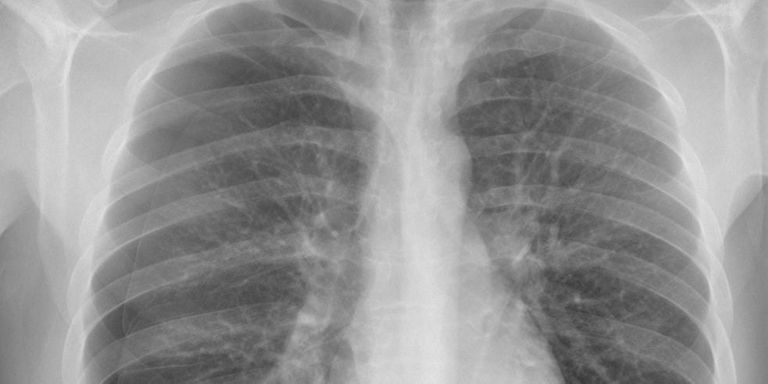
Pneumothorax is a critical medical condition, commonly referred to as a collapsed lung. It can be painful and is potentially deadly, so pneumothorax requires immediate attention. Pneumothorax may be caused by an underlying lung disease, a blunt chest injury or can occur after surgery. It can also occur spontaneously. It is generally diagnosed by radiologists through examining chest radiographs (chest x-rays) and can be difficult to confirm - can you see the pneumothorax in the image shown here?! For further information see the Radiopaedia definition or the Radiology Assistant.
Recently, the Society for Imaging Informatics in Medicine (SIIM) organized a Kaggle challenge to tackle the detection and segmentation of this condition on chest x-ray images. We are interested in creating an open-source algorithm that uses this Kaggle dataset (initially) and other open datasets as well as chest x-ray data we have collected at Radboud UMC to detect pneumothorax.
Research question and tasks
Detection and segmentation of pneumothorax in chest x-rays.
Tasks
- Research previous work on segmentation of 2D medical images
- Research previous work on pneumothorax detection
- Develop a deep learning method that tackles the task
- Issue late submissions in the leaderboard and compare model performance with winning submissions
- Test your model on data from different institutions
- Publish your algorithm under an open source license
Innovation
The goal is to integrate the algorithm in a set of chest x-ray analysis algorithms that are tested in routine clinical care at the Department of Radiology and Nuclear Medicine at Radboud UMC.
Requirements
- Students in the final year of a study in computer science, artificial intelligence, physics, or a related area.
- Knowledge of and experience with deep learning image analysis. You should sufficiently proficient to be able to implement systems described in this book.
Information
- Project duration: 6 months
- Location: Radboud University Medical Center
- For more information, please contact Keelin Murphy