Background
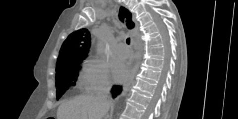
Osteoporosis is a silent age-related disease that slowly decreases the stability of the bones, which eventually leads to bones starting to fracture and collapse. These kind of fractures are referred to as compression fractures. Every year, millions of CT scans of the thorax and abdomen are made worldwide, for reasons other than osteoporosis and compression fractures, but providing the opportunity to detect compression fractures of the spine.
Research question and tasks
The goal of this project is to develop a system that automatically determines for each vertebra visible on a CT scan whether it is normal or whether it is fractured and collapsed, or is starting to collapse. For abnormal vertebrae, the system should quantify the degree to which the bone has collapsed in order to distinguish between early stage fractures and advanced fractures.
We have robust methods for automatically finding the spine in CT images and for delineating the individual vertebrae. A set of CT scans in which fractures have been labeled by a radiologist is available for this project. We also have access to a much larger set of CT scans in which the vertebrae have been manually delineated, but in which fractures have not yet been labeled. Part of the project will be to configure a workstation that can be used by radiologists to label additional data.
The resulting automatic fracture detection system will be a first step toward automatically reading all thorax CT scans in the background to detect fractures that would otherwise be missed. A mandatory part of the project is to make the developed algorithm available on the grand-challenge.org platform.
Requirements
- Student in the final year of a Master study in biomedical engineering, artificial intelligence, data science or similar
- Interest in medical applications and medical image analysis
- Good programming skills are important (Python)
- A solid theoretical understanding of deep learning is crucial, experience with deep learning frameworks (PyTorch) is beneficial
Information
- Project duration: 6 months
- Location: Radboud University Medical Center
- For more information, please contact Nikolas Lessmann. To apply for this project, please include your CV and a short summary of your background and your interest in this project in your email.